June 12, 2025
We are thrilled to announce the winners of the third annual LCIF Image Contest – 2025! We’re glad to continue celebrating the stunning microscopic images produced by the remarkable research within our facility and it would not be possible without your contributions. So, a heartfelt thank you to all participants for their outstanding submissions.
Here are the winners of the LCIF Image Contest 2025:
First-place winner
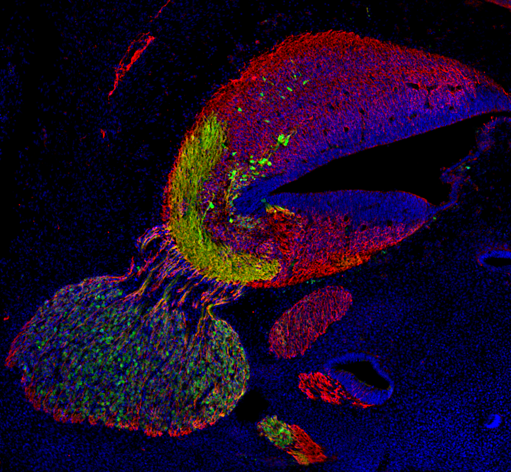
Immunolabeling of embryonic day 12 mouse embryo tissue reveals β3-tubulin⁺ (red)/α-synuclein⁺ (green) pioneer axons from the trigeminal ganglion (bottom left circular structure) weaving into the cerebellum, targeting α-synuclein⁺ neurons in the developing cerebellar nuclei.
Second-place winner
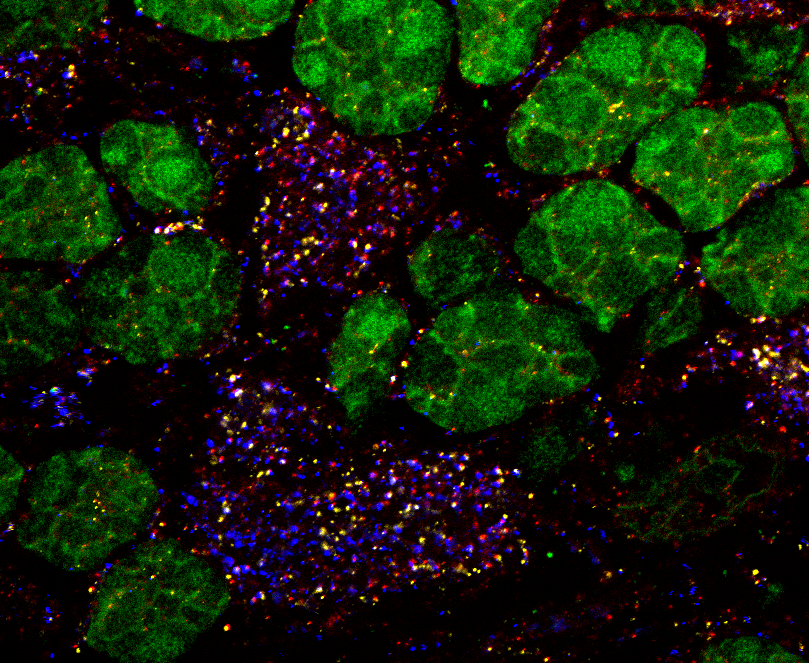
Acquired with Zeiss LSM 880 Confocal using expansion microscopy (physically enlarges tissue ~4× for higher resolution). Shows granule cells (green), presynaptic (red), postsynaptic (blue), and test protein (yellow) in sub-synaptic domains of cerebellar glomeruli.
Third-place winner
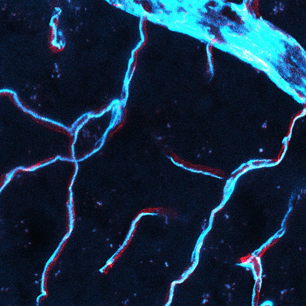
Z-stack projection of mouse brain cortex microvasculature using CD31 (red, endothelium) and CD13 (cyan, pericytes). A large vein connects to small capillaries. Pericyte labeling evokes a cloudy sky and lightning-like processes.
All entries (no order)
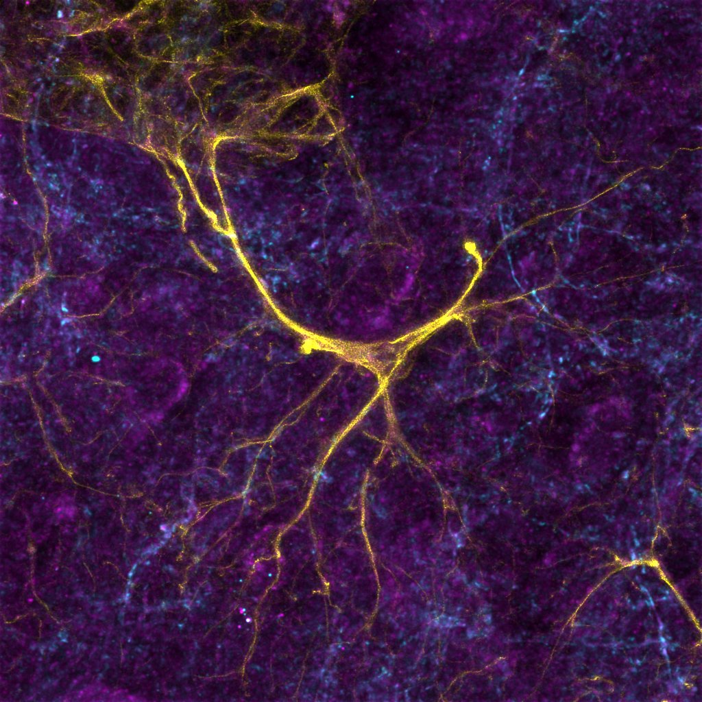
An astrocyte forms connections between vasculature and neurons in a mouse cortex. Astrocytes are labelled with anti-GFAP (yellow), GCaMP is labelled with anti-GFP (cyan) and P2Y1 receptors are labelled with anti-P2Y1 (purple).
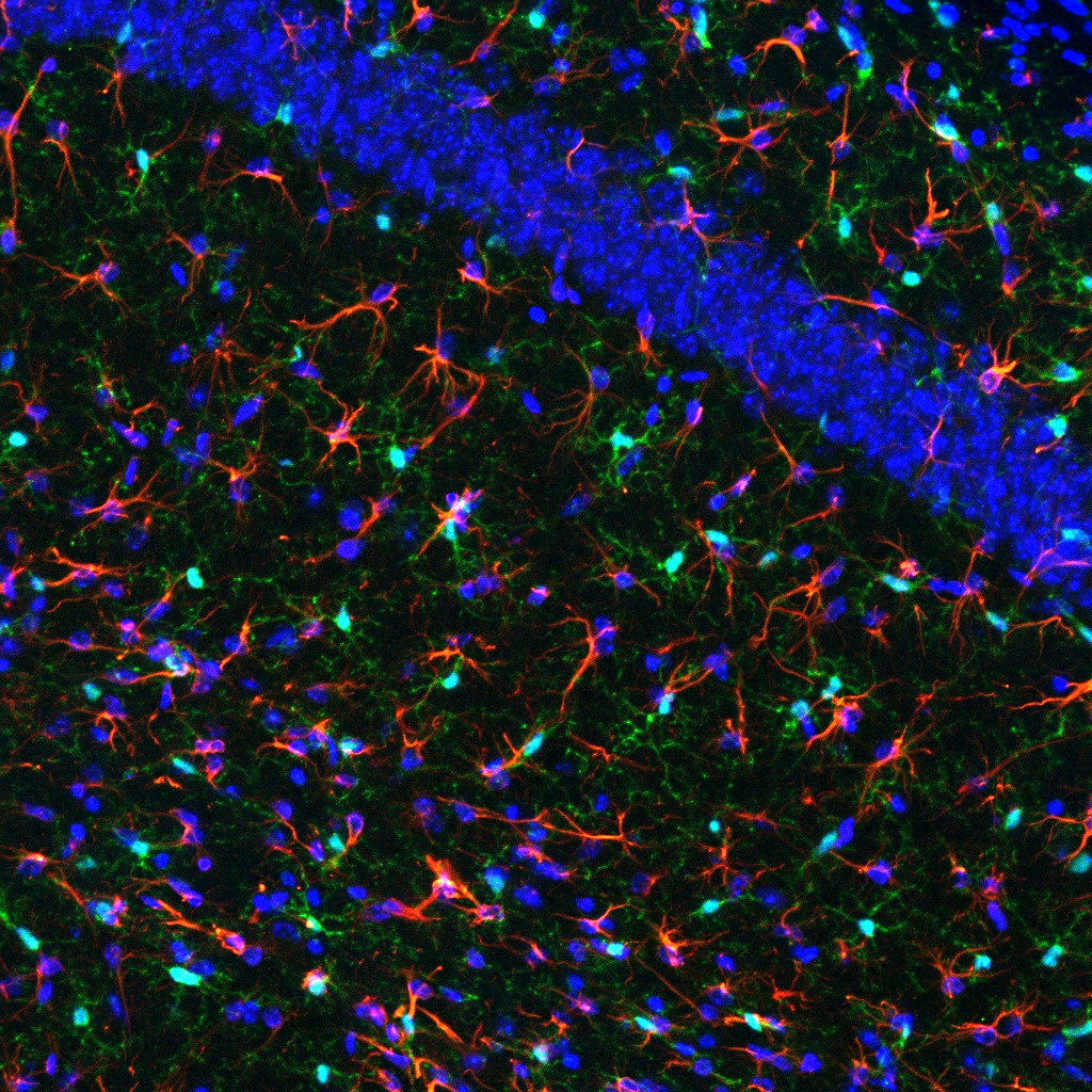
Microglia (green) and astrocytes (red) play essential roles in maintaining brain homeostasis and neuronal function, including immune response, blood–brain barrier maintenance and synaptic support. Section of a mouse hippocampus acquired with the Zeiss LSM880 microscope. Cell nuclei in blue.

A mesmerizing neuronal co-culture, visualized under fluorescence microscopy, evoking ethereal lightning storms within microscopic landscapes, harmonizing art with biology.
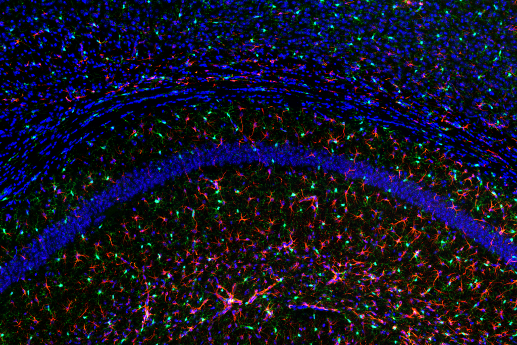
Astrocytes (red) and microglia (green), key players in brain homeostasis and immune response, labeled in a hippocampal section of a mouse exposed to amyloid beta oligomers. Tiled z-stack image acquired with the Zeiss LSM880 microscope. Cell nuclei depicted in blue.